Shrinking Lung Syndrome: A Case Report
DOI:
https://doi.org/10.56538/ramr.%20IWIE8726Keywords:
Shrinking lung syndrome , Systemic lupus erythematosus, Diaphragmatic dysfunction , Diaphragmatic ultrasound, restrictive pulmonary diseaseAbstract
Background: Shrinking lung syndrome (SLS) is a rare respiratory manifestation of systemic lupus erythematosus (SLE), characterized by dyspnea, diaphragmatic elevation, and a restrictive pattern on lung function testing, with no alternative pleuropulmonary disease accounting for these findings.
Objectives: To describe the case of a patient with recently diagnosed SLE who developed SLS, and to review the main pathophysiological, diagnostic, and therapeutic aspects.
Case report: We present the case of a 38-year-old woman with a history of smoking and hypertension who was recently diagnosed with SLE. She was evaluated with chest computed tomography, lung function testing (spirometry and lung volumes), and diaphragmatic ultrasound.
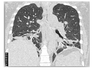
Results: The patient presented with a 5-month history of dyspnea, intermittent fever, polyarthralgia, and asthenia. Chest computed tomography revealed bilateral subsegmental atelectasis and elevation of the right hemidiaphragm. Lung function testing showed a severe restrictive pattern with markedly reduced diffusing capacity for carbon monoxide (DLCO), nd diaphragmatic ultrasound confirmed bilateral diaphragmatic paralysis. Following a diagnosis of SLS, treatment with oral corticosteroids and immunosuppression with cyclophosphamide was initiated, with a favorable clinical response.
Conclusions: SLS should be suspected in patients with SLE who present with dyspnea and an unexplained restrictive pattern. Its diagnosis is one of exclusion and requires a high index of suspicion. Diaphragmatic ultrasound is a useful tool for its assessment, and treatment with corticosteroids and immunosuppression is effective in most cases.