

Autor : Ortega, Mayra1, Gallego, Claudio1, Poropat Alejandra1, Salomone, CĂ©sar1
1Hospital General de Agudos Parmenio Piñero, Autonomous City of Buenos Aires, Argentina.
https://doi.org/10.56538/ramr.IJOE9390
Correspondencia : Mayra Ortega: mayra_ortega92@hotmail.com
ABSTRACT
Tuberculous pneumonia is a rare
disease, mostly described in patients with immuÂnosuppression or comorbidities
such as alcoholism or diabetes, with a radiological presentation similar to
bacterial pneumonia, which can lead to diagnostic delays. This study includes
patients diagnosed with tuberculosis and with a radiological image of
consolidation in lower lung fields, without associated comorbidities. Over the
course of 3 years, we identified 25 patients with these criteria among 628
evaluated cases of pulmonary tuberculosis. We didn’t find any relationship with
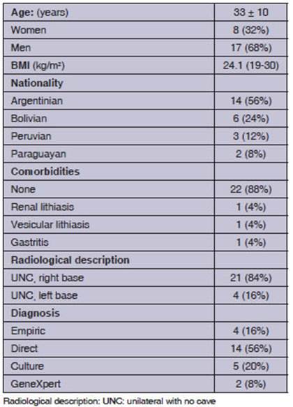
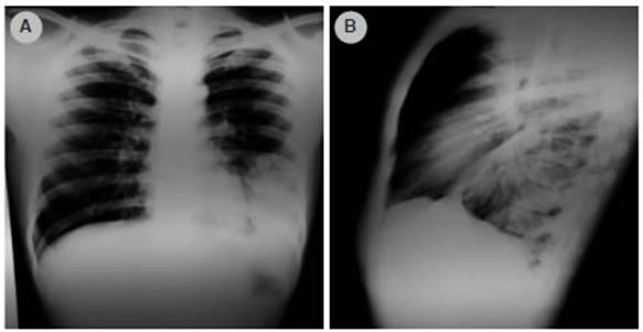
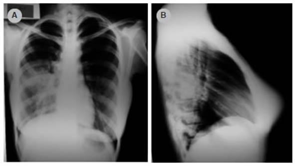
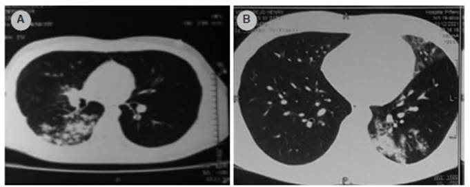
sex; and the right lower lobe was more frequently affected (84%) than the left.
Key words: Tuberculosis, Tuberculous pneumonia, Pneumonia
RESUMEN
La neumonía tuberculosa es una patología
poco frecuente, descripta mayormente en pacientes con inmunosupresión o
comorbilidades como alcoholismo o diabetes, con una presentación
radiológica similar a la neumonía bacteriana lo cual puede dar
lugar a retrasos en el diagnóstico. En este trabajo se incluyeron
pacientes con diagnóstico de tuberculosis e imagen radiológica de
consolidación en los campos pulmonares inferiores y sin comorbilidades
asociadas. En el transcurso de 3 años identificamos 25 pacientes con
estos criterios entre 628 casos de tuberculosis pulmonar evaluados. No
encontramos relación con el sexo, resultando más frecuente la
afectación del lóbulo inferior derecho (84%) que el izquierdo.
Palabras clave: Tuberculosis, Neumonía tuberculosa, Neumonía
Received: 07/29/2022
Accepted: 11/07/2022
INTRODUCTION
Tuberculosis is a public health
issue, caused by Mycobacterium tuberculosis. The World Health
Organization has been publishing reports since 1997 with the purpose of putting
an end to tuÂberculosis (TB) on a worldwide level.1,2
The “End TB” strategy proposes reducing the number of deaths by
95%, and the incidence by 90%, in order to achieve less than 10/100,000 inhabitÂants
in the 2015-2035 period.3 The Covid-19
pandemic threatens the established programs for TB, since it generated some
difficulties in access to healthcare.
Tuberculosis in Argentina is
still an important public health issue; in 2019 there was a reported rate of
27.8/100,000 inhabitants, 6.4% higher than in 2018 (26.2/100,000 inhabitants).
78% of new cases were of pulmonary localization.4
Primary tuberculosis develops in
patients who haven’t been previously exposed; it is common in pediatric
patients, and appears as a consolidaÂtion that affects the middle and lower
lobes and adjacent lymph nodes. Lower lung field TB, as referred to in the
reference studies, can be seen mainly in people living with HIV, diabetes,
renal or liver disease, and those receiving corticoids and diagnosed with
silicosis.5-7
This study was conducted for the
purpose of describing the epidemiological and radiologiÂcal characteristics of
tuberculous pneumonia in patients without immunosuppression showing
consolidation in the lower lung field.
MATERIALS AND METHODS
It is a retrospective (2017-2019
period) and prospective (2019-2021) study that analyzed TB cases treated at the
Hospital General de Agudos Parmenio Piñero within said periods. The
hospital is located in an area with high prevalence of tuberculosis (>100/100,000
inhabitants). The selection criteria for tuberculous pneumonia were: a)
positive bacilloscopy in sputum or bronchoalveolar lavage, or diagnosis of TB
with compatible epidemiology and cliÂnical symptoms, b) not having
comorbidities such as HIV, immunosuppression or addictions, c) not being
underweight (BMI <18.5), d) chest X-ray with image of consolidation in lower
fields.
The medical records of the
patients included serological testing for HIV, hepatitis B and C and VDRL
(Venereal DiÂsease Research Laboratory) test, the TB diagnostic method
performed and radiographic images.
Statistical analysis
Data obtained were analyzed with
descriptive statistics tools. Chi Square test was used for qualitative
variables (https://www.socscistatistics.com).
RESULTS
The analysis included 628 cases
of pulmonary tuberculosis, 25 of which (4%) were diagnosed as lower lung field
TB. 17 of those 25 cases (68%) were male. The mean age of patients was 33 ± 10
years. Most patients were Argentinian (56%), followed by Bolivians (24%),
Peruvians (12%) and Paraguayans (8%). The proportion of females wasn’t
significant, compared to males. Table 1 describes the characteristics of
patients being evaluated. There is predominance of right lower lobe
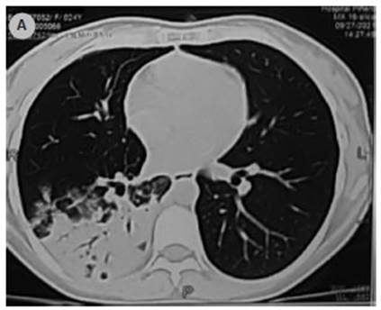
involvement: 84%. Figures 1 to 4 show examples of the images found.
DISCUSSION
In the group admitted
to the study, we identified 4% of patients with lower lung field tuberculosis
without associated comorbidities; there were no significant differences
regarding sex; and involveÂment of the right lung base was predominant in the
X-ray.
One of the published
studies describing 62% predominance of women suggests the hypothesis that women
have intercostal breathing with less diaphragmatic stretching that could result
in less ventilation and less expansion of the lower lobes.8 In our
case report we didn’t find any differences relating to sex that support such
hypothesis.
The observed
predominance of the right lung base coincides with what was evidenced in previÂous
studies.8,9 In the studies of India (61%) and Taiwan (64%), a
predominance of the right lung base involvement was found that was close to the
one found by us (84%). The hypothesis that was suggested for this finding is
that the main right bronchus is anatomically shorter and has a sharper angle
compared to the left bronchus, thus the infectious microorganisms more easily
propagate towards the right lower lobe.8-10
Many authors have
described that lower lung field TB occurs more frequently in specific groups of
patients with diseases without immunosuppresÂsion.10-11 In agreement
with this, some studies in India found that tuberculous pneumonia is more
frequent in: diabetes (29%), patients living with HIV (12%), patients receiving
treatment with corticosteroids (12%), with liver disease (11%), and with renal
disease (5%). On the other hand, we couldn’t find any published studies that
describe this form of TB manifestation in groups of patients without
comorbidities.8
Lower lung field TB
is an atypical presentation of pulmonary TB. The radiographic image similarÂity
with acute community-acquired pneumonia or even some types of bronchogenic
adenocarcinomas entail diagnostic delays.12 One of the proposed
explanations is the transbronchial perforation of an affected hilar lymph node,
with dissemination to the adjacent parenchyma.13
One limitation of
this study is the lack of data identifying the exact time of diagnostic delay
of the described group of patients. One strength is the fact of having shown
that tuberculous pneumonia occurs even in patients without comorbidities
considered as predisposing factors.
To conclude,
tuberculosis must be included in the group of differential diagnoses of
patients who show consolidation of the lower lung field and have a history of
exposure or epidemiological risk, even if they don’t have significant comorÂbidities
or immunosuppression, thus avoiding diagnostic delay.
Conflict of interest
There is no conflict
of interest.
REFERENCES
1. Global tuberculosis report
2020.
https://apps.who.int/iris/bitstream/handle/10665/336069/9789240013131-eng.pdf
2. Organización Mundial de la Salud. Informe mundial de tuberculosis 2020.
cms-decommissioning (who.int)
3. Raviogli.M. The
end TB strategy. World Health OrganizaÂtion. Disponible:
https://www.who.int/publications/i/item/ WHO-HTM-TB-2015.19.
https://bancos.salud.gob.ar/sites/default/files/2021-03/boletin-epidemiologico-tuberculoÂsis-2021.pdf
4. Boletin N° 4
Tuberculosis en la Argentina.2021. MinisteÂrio de Salud Argentina (marzo 2021).
Disponible:
https://bancos.salud.gob.ar/sites/default/files/2021-03/boletin-epidemiologico-tuberculosis-2021.pdf
5. Anton P, Jhon B, Elinor L. Manifestaciones
clínicas y comÂplicaciones de la tuberculosis pulmonar. 2021. Uptodate. (noviembre del 2021). Disponible en:
https://www.uptodate.com/contents/clinical-manifestations-and-complications-of-pulmonary-tuberculosis?search=lower%20lung%20fied%20tuberculosis§ionRank=1&usage_type=defa
ult&anchor=H17&source=machineLearning&selectedTitle=1~1&display_rank=1#H1
6. Castiñera
A, López MR, Peña MJ, Liñares M.
Manifestación radiológicos de la tuberculosis pulmonar. Med Integral. 2002;39:192-206.
7. Robert H, Fordham
V, Elinor L. Epidemiologia de la tuÂberculosis. Uptodate. 2021. Disponible en:
https://www.uptodate.com/contents/epidemiology-of-tuberculosis
8. Singh SK, Tiwari
KK. Clinicoradiological Profile of Lower Lung Field Tuberculosis Cases among
Young Adult and Elderly People in a Teaching Hospital of Madhya Pradesh, India.
J Trop Med. 2015;2015:230720. https://doi.org/10.1155/2015/230720
9. Lin CH, Chen TM,
Chang CC, Tsai CH, Chai WH, Wen JH. Unilateral lower lung field opacities on
chest radiography: a comparison of the clinical manifestations of tuberculosis
and pneumonia. Eur J Radiol. 2012;81:e426-30. https://doi. org/10.1016/j.ejrad.2011.03.028
10. Gutierrez J,
Zamudio S. Neumonía tuberculosa. Reporte de 20 casos y estudio caso
control. Acta Med Peru 2011;18:5-11. https://pesquisa.bvsalud.org/portal/resource/pt/lil-506725
11. Ajai K, Saurabh
S. Lower lung field pulmonary tuberÂculosis: An overview. Indian.2015. Indian Acad Clin Med.
Disponible:
https://www.researchgate.net/publication/282282391_Lower_lung_field_pulmonary_tuberculoÂsis_An_overview
12. Lee K, Choe H,
Kim S. Clinical investigation of caviÂtary tuberculosis and tuberculous
pneumonia. Korean J Intern Med.
2006;21:230-5.
https://doi.org/10.3904/kjim.2006.21.4.230
13. González A, Fernández Cáceres M,
Baldini M, Monteverde A. Tuberculosis pulmonar de campos inferiores. Medicina
(B. Aires)

| GalerĂa de imágenes | ||
| Mujer joven con afectaciĂłn pulmonar bilateral y alteraciĂłn de la conciencia | ||
Autores: Churin Lisandro |
 |
|